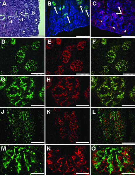
Figure 1. Renal morphology and immunohistochemistry - Early third trimester fetal monkey kidney and nephrogenic zone.
(A) The fetal monkey kidney in the early third trimester shows a clear nephrogenic zone in the cortical region with the ureteric bud (arrowhead) and the metenephric mesenchyme (arrow) evident. The four stages of developing glomeruli are also present and represent the vesicle (a), S-shape (b), capillary (c), and mature (d) glomerulus. Hematoxylin and eosin (H&E). (B-C) The nephrogenic zone is shown with the nuclei stained with DAPI (blue), metanephric mesenchyme (arrowheads), developing podocytes, and mature podocytes (arrows) positive for synaptopodin (green) and nestin (red). (D-I) Mature podocytes in terminally differentiated glomeruli are positive for nestin (red) and synaptopodin (green). (J-O) The endothelial marker CD31 (green) is not evident with the nestin positive cells (red) in these specimens (A-C, 20x, bar = 100 μm; D-F, 40x, bar = 50 μm; G-I, 100x, bar = 20 μm; J-L, 40x, bar = 50 μm; M-O, 100x, bar = 20 μm).

