
Figure 2. Morphology of cultured renal cortical cells.
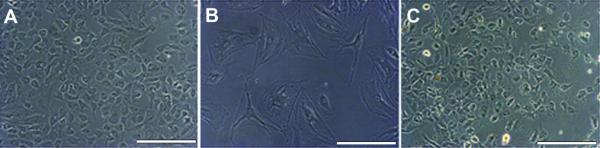
(A) Through passage 4, all substrates studied showed cells with the “cobblestone” appearance suggestive of undifferentiated podocytes. (B) At passage 5, cultures contained cells that had a differentiated podocyte cell morphology with arborization, processes extended from cell bodies, and a large cytoplasmic to nuclear volume ratio. (C) Renal ECM contained cells with an undifferentiated podocyte morphology through all passages (1 through 5) (20x, bar = 100 μm).