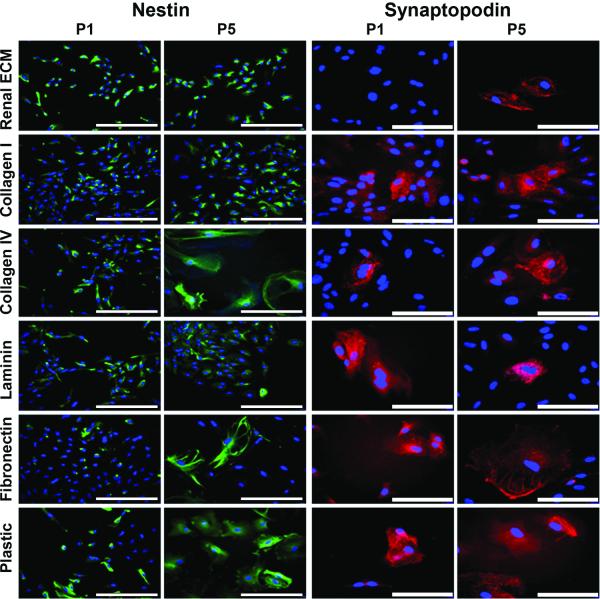
Figure 4. Immunocytochemistry - Cultured cortical cells.
Cells from passage 1 (P1) grown on each of the substrates included nestin positive cells (green) (20x, bar = 100 μm). Nuclei stained with DAPI (blue). At passage 5 (P5), nestin positive cells decreased on all substrates except renal ECM and collagen I. Cells from P1 cultured on five of six substrates contained very few cells that were synaptopodin positive (red, 40x, bar = 50 μm). None of the cells grown on renal ECM showed evidence of synaptopodin expression. At P5, there was an increase in synaptopodin positive cells suggesting a more mature podocyte phenotype.

