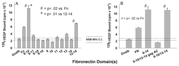
Figure 3.
Mapping the VEGF-binding site on C-terminal Fn. A and B, 125I-VEGF was incubated for 2 hours on wells coated with either individual or continuous rFn type III domains (0.2 μmol/L) (A) or wells coated with rFn molecules encompassing both the integrin (9 to 10) and heparin-binding domains (12 to 14) (B). GagAC denotes mutation of the heparin-binding domains on type III module 13 and 14. After washing, bound VEGF was eluted with 0.1 mol/L NaOH/1% SDS and radioactivity quantified in a gamma counter. Data are the means±SEM of 3 to 4 independent experiments performed in triplicate. Dotted lines are 95% confidence intervals for control binding to albumin.

