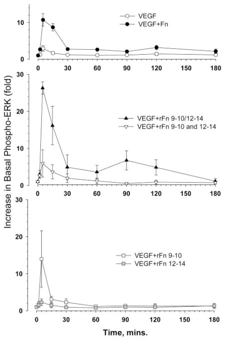
Figure 6.
HUVEC Erk phosphorylation in response to VEGF and Fn moieties. HUVECs were exposed to 10 ng/mL of VEGF165, combined with the indicated Fn proteins (0.2 μmol/L). At the time points, cells were harvested and processed to measure phospho-Erk and total Erk protein by Western blotting. Each point on the graph is the mean of data obtained from 4 to 5 separate experiments and quantified as described in Materials and Methods.

