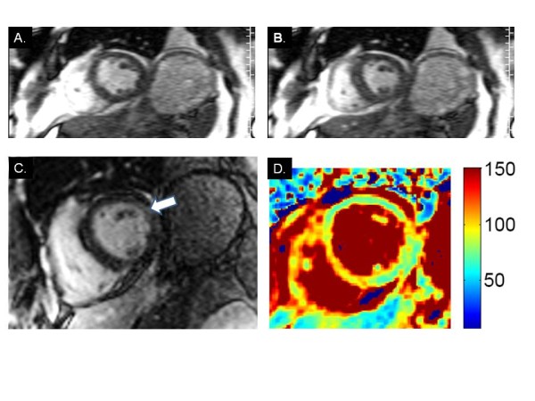
Figure 1.

CMR findings in wet beriberi. End-diastolic (A) and end-systolic (B) frames from short axis cine imaging demonstrate global hypokinesis of the left ventricle. (C.) LGE image shows borderline hyperenhancement of the basal lateral wall (arrow). (D) T2 mapping demonstrates significantly elevated myocardial T2 values suggestive of diffuse edema.
