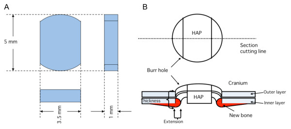
Figure 1.
Three-dimensional views of a hydroxyapatite ceramic button (HAP) and the implantation of HAP samples into bone defects (burr holes). A; HAPs were made for cranial repair (cranioplasty) in rats. Both sides of the HAP were cut in order for bone formation to extend into the space around the bone defect. B; The panel demonstrates how bone formation was measured. Bone formation was quantified by measuring the length of new bone extension into the inside of the bone defect and the thickness of the edges of the bone defect.

