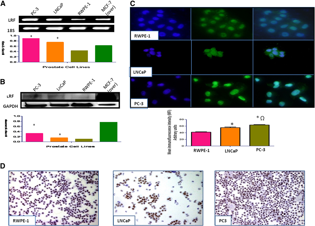
Figure 1. Expression of LRF/Pokemon in human prostate cell lines.
LRF mRNA transcripts (A) and protein expression (B) in prostate cell lines with densitometric analysis indicate that cancer cell lines have significantly higher expression than normal prostate cell lines. MCF-7 cells overexpressing LRF were used as a positive control. (C) Immunohistochemical expression of LRF in prostate cell lines. DAPI (blue) was used as nuclear counterstain (200x–400x). (D) Immunohistochemical expression of LRF in prostate cell lines. DAB was used as a chromogen and hematoxylin as counterstain (200x–400x).

