Figure 2.
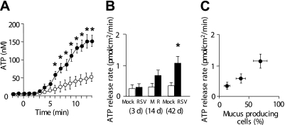
ATP release rates from cultures after RSV infection. (A) ATP accumulation in airway surface liquid when epithelial cell surface-mediated ATP hydrolysis was inhibited by the addition of ecto-ATPase inhibitors (β, γ-methylene-ATP [300 μM], ebselen [30 μM], and levamisole [10 mM]) to airway surface liquid at t = 0. Open circles and solid circles indicate mock- and RSV-infected cultures, respectively, at 42 days after infection. (B) ATP release rates in resting cultures at 3, 14, and 42 days after RSV (or mock) infection, as measured from ATP accumulation rates by using the method depicted in Fig. 2A and Ref. 12. (C) Correlation between goblet cell percentages (data imported from Fig. 1D) and ATP release rates (data imported from Fig. 2B). *Significant difference (P < 0.05) over mock-infected control cultures. Values are mean ± SE of four Transwells/subject established from three different subjects.

