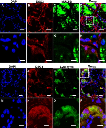
Figure 5.
Immunofluorescent double labeling of DSG3 and MUC5B or lysozyme in human sinus SMGs. (A–H) Double-labeling of DSG3 and MUC5B. (I–P) Double labeling of DSG3 and lysozyme. Labeling by the DSG3 antibody was detected with a rhodamine-labeled secondary antibody. Labeling of MUC5B and lysozyme were detected with FITC-labeled secondary antibody in the same section. The overlap of DSG3 (red) and MUC5B or lysozyme (green) labeling appeared orange (Merge). Double labeling revealed colocalization of DSG3 and lysozyme in the same SMG cells, but no colocalization of DSG3 and MUC5B. No staining was evident under negative control conditions. Scale bars, 20 μm in A–D and I–L; 10 μm in E–H and M–P. The images in the white dotted box in D and L are magnified and shown in E–H and M–P, respectively.

