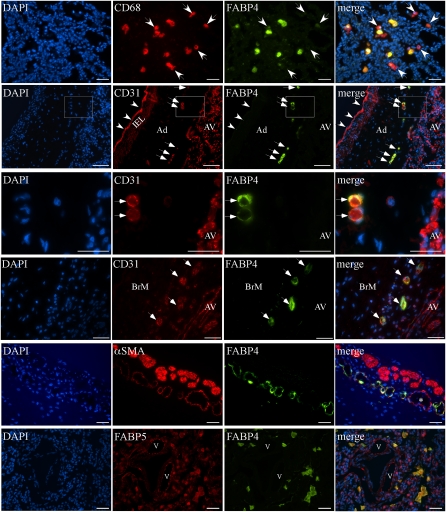
Figure 3.
Double immunofluorescence analysis for FABPs, CD31, CD68, and α-smooth muscle actin (αSMA) on baboon and human BPD sections. Representative images are shown. First row (baboon BPD): FABP4 is colocalized with CD68 in most, but not all, alveolar macrophages. White arrows indicate CD68-positive, but FABP4-negative, cells. Second row (human BPD): CD31 and FABP4 are colocalized in vasa vasorum ECs in the adventitia (Ad) of a pulmonary artery (white arrows), but not in pulmonary arterial ECs (white arrowheads) or alveolar vessels (AV). Internal elastic lamina (IEL) demonstrates autofluorescence. Third row: Higher magnification of the boxed area in row 2 demonstrates CD31 expression in alveolar vasculature and vasa vasorum ECs, but FABP4 expression only in vasa vasorum ECs (white arrows). Fourth row (human BPD): CD31 and FABP4 are colocalized in small vascular ECs in the bronchial mucosa (BrM, white arrows). Fifth row (human BPD): αSMA, a marker of mature pericytes and smooth muscle cells, is detected around some FABP4-positive peribronchial vessels. Larger vessels with continuous coverage of αSMA-expressing cells do not express FABP4 (asterisk). Sixth row (human BPD): In contrast to the widespread expression pattern of FABP5 in several cell types, including alveolar epithelial and endothelial cells lining pulmonary vessels (v) in the lung parenchyma, FABP4 is detected only in FABP5-positive alveolar macrophages. Scale bars = 25 μm.

