
Figure 1.
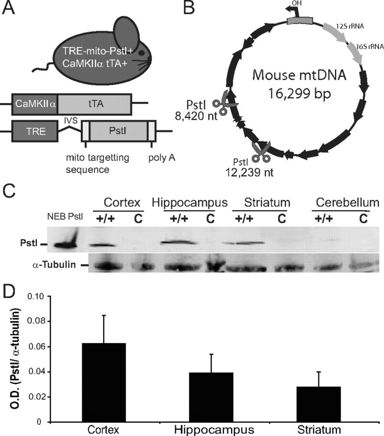
Mito-PstI mouse expresses mitochondrial-targeted restriction endonuclease in the CNS. A, Schematic representation of the experimental mito-PstI mouse and the transgenic constructs it harbors. B, Representative mtDNA map showing the targeted sites of PstI at 8420 and 12,239 nt (scissors). Black arrows denote protein-coding genes. C, Western blots of brain lysates using antibodies probing for mito-PstI protein expressed in four regions of the CNS (cortex, hippocampus, striatum, and cerebellum) of 2-month-old mito-PstI (+/+) or control (c) animals. α-Tubulin immunoreactivity was used to ensure equal protein loading. D, Quantification of the optical density (O.D.) of immunoblotted PstI protein levels from mito-PstI mice. Results are normalized against α-tubulin immunoreactivity. Values are mean ± SEM (n = 3, NS).