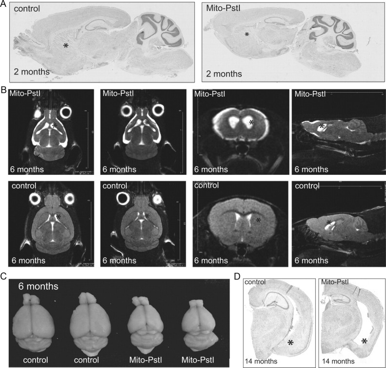
Figure 3.

Mito-PstI mice have an age-related neurodegeneration with a preferential degeneration of the striatum. A, Parasagittal Nissl-stained histological sections of 2-month-old animals. B, Nuclear magnetic resonance imaging of 6- to 7-month-old control and mito-PstI mice in vivo reveal ongoing degeneration with ablation of the striatum with cortical atrophy. The striatum is denoted by asterisks. White areas represent CSF (n = 2/group). C, Gross morphology reveals abnormal brain size and appearance of mito-PstI animals as compared to control at 6–7 months of age. Control brain weight was ∼0.42–0.45 g, and mito-PstI weight was ∼0.27–0.30 g. D, Nissl staining of coronal sections reveals that, at 14 months of age, degeneration in the mito-PstI occurs in the ventral aspects of the striatum (denoted by asterisks), which is absent in controls.
