
Figure 2.
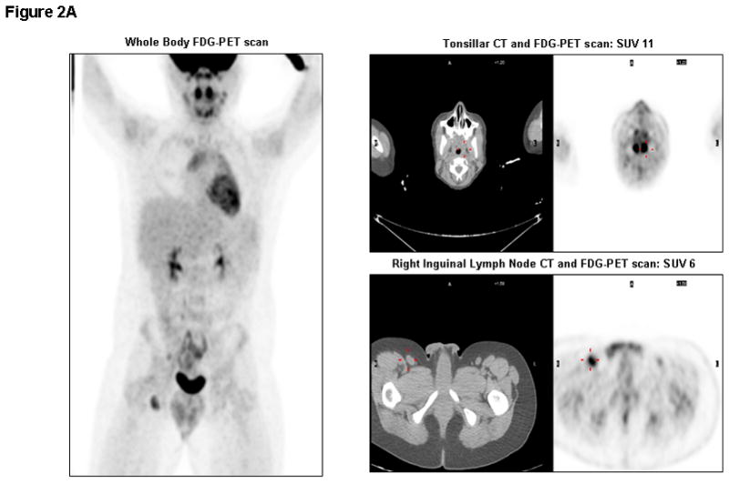
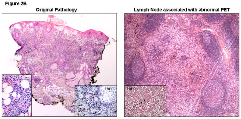
Figure 2A. Whole Body FDG-PET/CT scan on day +237: Significantly elevated metabolic activity in bilateral tonsils (SUV 11) and right inguinal lymph nodes (SUV 6), in addition to cervical (SUV 4.5), mediastinal (SUV 5), and mesenteric (SUV 3.6) lymph nodes. Corresponding CT images of tonsils and inguinal lymph node are shown with abnormal signals marked in red.
Figure 2B. Skin Biopsy at Original Presentation: Histopathological examination of a lesion on the face showed acute spongiosis with prominent vesiculation and necrosis, as well as a dense superficial and deep infiltrate with adnexal involvement and extension into the subcutaneous fat. Left inset demonstrates an infiltrate composed of large, atypical angulated lymphocytes. Right inset reveals diffusely EBV positive lymphocytes on EBERish stain (hematoxylin and eosin, 20X; insets, original magnification 400X).
LN Biopsy post-CTL Infusion (Day+243): Histopathological examination of the inguinal lymph node showed preserved architecture and reactive changes, in the form of nodular paracortical hyperplasia (dermatopathic changes), follicular hyperplasia and sinus histiocytosis. A rare EBV positive small lymphocyte (inset, arrow) was seen (hematoxylin and eosin, 40X; inset, EBERish for EBV, original magnification 400X). EBERish: in situ hybridization for EBV encoded small RNAs.