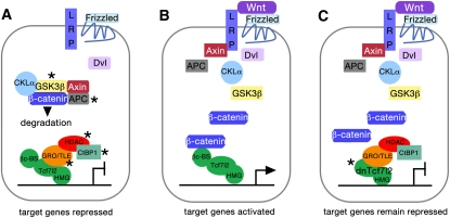
Figure 1.
(A–C) Schematics of Wnt signaling components in the absence of a Wnt signal (A), the presence of a Wnt signal (B), and the presence of a Wnt signal blocked by the displacement of Tcf7l2 by dnTcf7l2 (C). (A) Without a Wnt signal, β-catenin is phosphorylated by a protein complex surrounding GSK3β and sent for ubiquitination. In the nucleus of the cell, Tcf7l2 associates with the Groucho-TLE corepressors, as well as other repressors (HDAC and CtBP1). Transcription of Wnt target genes is repressed. Asterisks indicate components of the Wnt signaling pathway that Vacik et al. (2011) found are up-regulated by Vax2 (see also C). (B) The Wnt ligand binds to its Frizzled receptors and LRP coreceptors. Dvl breaks up the GSK3β-centered protein complex, releasing β-catenin, which translocates to the nucleus, displaces corepressors, and attaches to the β-catenin-binding site (βc-BS) on Tcf7l2. Target Wnt genes are expressed. (C) DnTcfl72 has displaced Tcf7l2 in the nucleus. Because dnTcfl72 lacks a β-catenin-binding site, it remains a transcriptional repressor even in the presence of a Wnt signal and the subsequent availability of β-catenin. The cell has, in effect, become “blind” to the Wnt signal (see Vacik et al. 2011).

