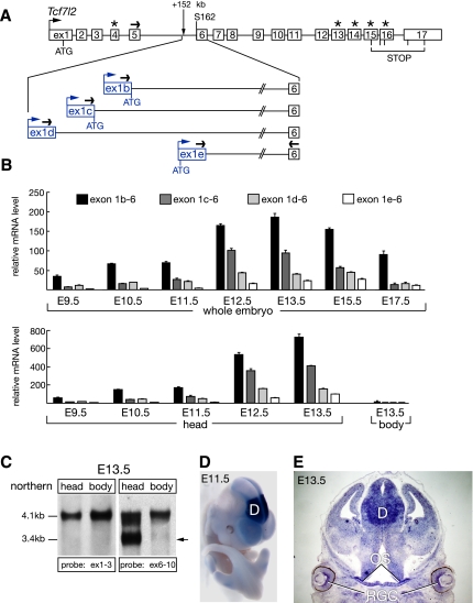
Figure 2.
Truncated Tcf7l2 mRNAs originate in Tcf7l2 intron 5. (A) 5′ RACE reveals the existence of four alternative Tcf7l2 first exons in Tcf7l2 intron 5. Exons 1–17 of the mouse Tcf7l2 gene are shown in white (Weise et al. 2010). Alternative exons 4, 13, 14, 15, and 16 are marked with asterisks. Alternative first exons of the truncated Tcf7l2 mRNAs are shown in blue. Black arrows depict primers used in the qRT–PCR experiments, and blue arrows depict alternative TSSs for exons 1b–e. (B) qRT–PCR analysis of the expression of the alternative Tcf7l2 mRNAs 1b, 1c, 1d, and 1e during embryogenesis, relative to Hprt. Error bars are mean ± SD (n = 3). (C) Northern analysis of Tcf7l2 mRNAs at E13.5. (Left two lanes) A probe against exons 1–3 detects only the full-length Tcf7l2 mRNAs (∼4.1 kb) in both the E13.5 head and body. (Right two lanes) A probe against exons 6–10 detects the full-length mRNAs in both the head and body, and also the truncated Tcf7l2 mRNAs (∼3.4 kb, arrow), but only in the E13.5 head. (D) Whole-mount in situ hybridization with a probe against exon 1b reveals a strong diencephalic expression (D) of the alternative Tcf7l2 mRNA 1b at E11.5. (E) In situ hybridization on a coronal section through an E13.5 head reveals the expression of the Tcf7l2 mRNA isoform 1b in ganglion cells of the developing retina (RGC), the optic stalk (OS), and diencephalon (D).

