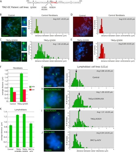
Figure 7.
DC patient cells harboring TIN2 mutations suffer loss in sister telomere cohesion. (A) DC mutations in patient cell lines are indicated. (B–E) Loss in sister telomere cohesion in DC patient fibroblasts harboring the TIN2p.Q269X mutation. FISH analysis with a 16ptelo (B,C) or 10cen (D,E) probe. DNA was stained with DAPI (blue). Bar, 5 μm. Histograms showing the distance between FISH signals (Table 3) are on the right, with the average (Avg) distance ± SEM indicated. (F) Graphical representation of the average distance ± SEM. (G–K) Loss in sister telomere cohesion in DC patient LCLs harboring the following mutations: TIN2p.K280Rfs36X, TIN2p.R282H, and DKC1p.A2V. (G–J) FISH analysis with a 16ptelo probe. DNA was stained with DAPI (blue). Bar, 5 μm. Histograms showing the distance between FISH signals (Table 3) are shown on the right, with the average (Avg) distance ± SEM indicated. (K) Graphical representation of the average distance ± SEM.

