Figure 2.
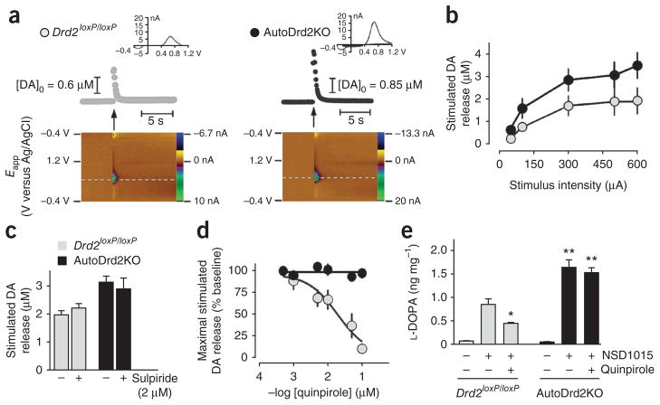
Increased DA release and DA synthesis in autoDrd2KO mice. (a) DA release in the dorsal striatum evoked by a single stimulus pulse (300–600 μA, 0.6 ms per phase, biphasic; arrows). Top, time course of DA concentration changes. Insets represent the background-subtracted cyclic voltammograms indicative of DA. Bottom, two-dimensional representations of the voltammetric data. The voltammetric current is plotted against the applied potential (Eapp) and the acquisition time. (b) Input-output relationship of DA release elicited by single-pulse stimulation across a range of stimulus intensities in the dorsal striatum of Drd2loxP/loxP (n = 4) and autoDrd2KO mice (n = 5) (F1,29 = 10.27, P < 0.001). (c) Stimulated DA release in autoDrd2KO (n = 16) and control mice (n = 11) does not change in the presence of 2 μM sulpiride (Drd2loxP/loxP mice, n = 8; autoDrd2KO mice, n = 7). (d) Effect of quinpirole on electrically stimulated DA release (F5,34 = 17.94, P < 0.001). (e) Tyrosine hydroxylase activity assessed by L-DOPA accumulation in striata of Drd2loxP/loxP and autoDrd2KO mice receiving saline or 100 mg per kg, intraperitoneal, of NSD1015. Quinpirole (0.5 mg per kg, intraperitoneal) was given 30 min before NSD1015 (two-way ANOVA genotype × treatment interaction: F2,17 = 8.58, P < 0.005; treatment: F2,17 = 48.15, *P < 0.05 between NSD1015 treated mice receiving or not receiving quinpirole; genotype: F1,17 = 34.84, **P < 0.001, post hoc Fisher analysis). Error bars represent s.e.m.

