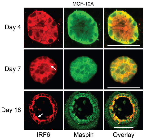
Fig. 4.
In vitro confirmation of interferon regulatory factor 6 (IRF6) lumenal localization. MCF-10 A cells were grown in a 3-D culture system that allows for the growth of hollow, lobular acini in cell culture. Acini were analyzed at three stages of growth: day 4 (immature, unorganized acinus), day 7 (partially organized acinus; hollowing begins via apoptosis of inner cells), and day 18 (acinus is mature and completely hollowed). IRF6 immunofluorescence is shown in red and Maspin immunofluorescence is shown in green. The overlay shows both Maspin and IRF6 with co-localization of these proteins highlighted by yellow. Arrows depict the lumenal localization of IRF6. The assay was carried out according to the protocol by Debnath et al. (Debnath et al. 2003). Imaging was carried out on a Zeiss 510 META confocal laser scanning microscope. Bar represents 50 μm.

