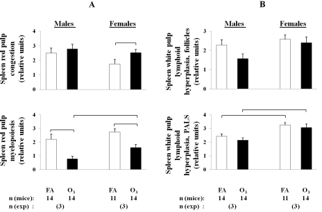
Figure 6.
Changes in leukocyte populations in the spleens of ozone-exposed and FA-exposed male and female mice after K. pneumoniae infection. After ozone or FA-exposure, mice were infected with K. pneumoniae as described in the legend of Figure 2, and the spleens were sampled and analyzed for histopathologic changes in both the red and white pulp. The myeloid compartment of the red pulp and the follicular and PALS compartments of the white pulp were each individually semiquantitatively scored for hyperplasia of the relevant leukocyte populations on a 0-4 numerical scale: 0 for normal, 1 for minimal hyperplasia, 2 for mild, 3 for moderate, and 4 for severe florid hyperplasia. Acute congestion of the red pulp was graded by a similar scale. A: Comparison of histopathologic changes in the red pulp in males versus females in response to ozone-exposure and pneumonia. B: Comparison of histopathologic changes in the white pulp in males versus females in response to ozone-exposure and pneumonia. The number of independent experiments was 3 for each sex, and the total number of mice is shown in the bottom of the Figure. Statistical analysis of data was performed using a t-test. Differences were considered significant if p < 0.05 and are indicated with the brackets.

