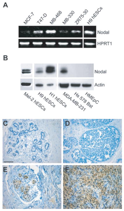
Fig. 6.
Nodal Expression Correlates with Breast Carcinoma Progression. A) Semiquantitative RT-PCR analysis of Nodal, mRNA expression in: MCF7, T47D, MDA-MB-468, MDA-MB-330, and ZR75-30 (human breast carcinoma cells). H9 hESC mRNA is used as a positive control for gene expression and HPRT1 is used as a loading control. B) Western blot analysis of Nodal in: MEL-2, H1 and H9 human embryonic stem cells (hESCs); MDA-MB-231, human metastatic breast carcinoma cells; Hs 578 Bst normal human myoepithelial cells; and HMEpC normal human mammary epithelial cells. Actin is used as a loading control. C) Immunohistochemical analysis of Nodal staining (dark color) in normal breast tissue, ductal carcinoma in situ (DCIS), nonspecific invasive ductal carcinoma (IDC) and metastatic IDC (to lymph nodes). The expression and prevalence of Nodal staining in breast tissue was designated as none, weak (<25%), moderate (25–75%) or strong (>75%). Spearman’s rank correlation showed a significant positive correlation between breast cancer progression and Nodal expression (p < 0.05). Bar equals 100 μm for all representative images shown.

