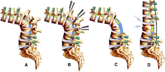
Fig. 2.
VCD diagrams. a Pedicle screws were inserted before the osteotomy was performed. b A high-speed drill was used to decancellate the deformed vertebrae. c Many posterior elements were removed along with the residual disc. d Postoperative lateral view shows that correction is achieved by elongating and opening (arrow) the anterior column and shortening the posterior column, and the residual bone takes the place of metal mesh described in the VCR technique, serving as a “bony cage”

