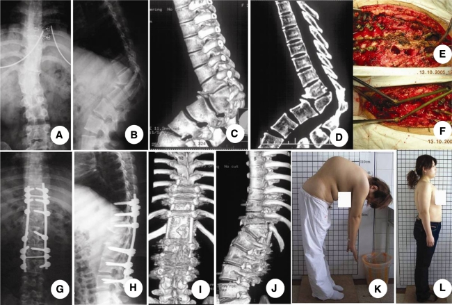
Fig. 3.
A 24-year-old patient with Pott’s disease. The patient’s main complaints were low back pain and cosmetic issues. a–d Preoperative X-ray and CT scan reconstruction showed a sharp angle in thoracolumbar spine. e, f Intraoperative picture of a vertebral column decancellation (VCD) was taken at T12 and L1. The VCD was begun with the probe of the pedicle of deformed vertebral body. A high-speed drill was used to enlarge the two pedicle holes. The residual upper and lower cartilaginous endplates and discs, posterior walls of the vertebral body and posterior elements were removed carefully, followed by cantilever technique and pedicle instrument; g, j 3 years’ follow-up X-ray and CT scan show a solid fusion between T12 and L1. k, l Preoperative and postoperative clinical pictures show the improved cosmesis

