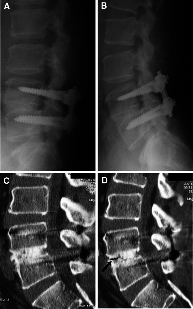
Fig. 3.

Imaging studies obtained in the illustrative case. Postoperative flexion (a) extension (b) lateral radiograph obtained 1 year after surgery. These radiographs demonstrating fusion were achieved at the L4/5 level. c and d Postoperative flexion and extension CT scan taken 1 year after surgery. Flexion CT scan (c) showing that fusion was complete at the L4/5 level. However, extension CT (d) showed a gas pattern in the interbody space. We then judged that successful arthrodesis had not been achieved
