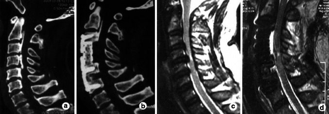
Fig. 1.
Case 1. Preoperative CT image (a) shows a mixed type OPLL and postoperative CT image (b) shows complete resection of OPLL using anterior corpectomy and fusion. Preoperative sagittal MRI of the cervical spine (c) shows signal intensity at C4–5 and postoperative sagittal MRI (d) shows the area of signal intensity decreased using anterior decompression

