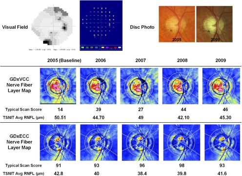
Figure 3.
A glaucomatous eye falsely identified as having apparent RNFL loss over time by GDxVCC, despite nonprogressing visual fields (top left) and optic disc stereophotographs (top right). On the detection software map (Progressor Medisoft, London, UK), each bar represents one test with the bar length showing to the depth of the defect and the color showing the P value of the regression slope. None of the locations demonstrated progression in this patient. The baseline GDxVCC (middle) image (TSS = 14) demonstrated ARP, and during 4 years of follow-up, there was a 32-unit increase in TSS and a 5.19-μm reduction in the TSNIT average RNFL thickness. The baseline GDxECC image (bottom) demonstrated a normal retardance pattern (TSS 91), and the absolute change in TSS was 7 units with less fluctuation in RNFL thickness.

