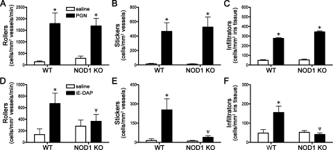
Figure 4.
Differential functions for NOD2 and NOD1 in PGN-triggered uveitis. NOD1 KO mice or WT controls were administered an i.v.t. injection of 5 μg PGN or saline, and ocular inflammation was assessed by intravital microscopy 6 hours later. The number of rolling (A), sticking (B), and infiltrating (C) leukocytes within the iris was quantified. Values are mean ± SE (n = 8–12 mice per group). * P< 0.05 saline vs. PGN within a genotype; ψP < 0.05 KO vs. WT mice treated with PGN. (D–F) Quantification of rolling, sticking, and infiltrating leukocytes within the iris in response to 100 μg iE-DAP i.v.t. injection. * P < 0.05 saline vs. iE-DAP within a genotype; ψP < 0.05 KO vs. WT mice treated with iE-DAP.

