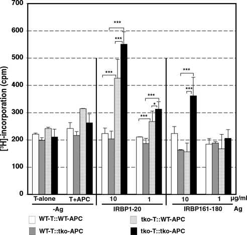
Figure 3.
IRBP-specific T cells are present in naive TAM tko mice. The T cells (4 × 10⁴) used for each [3H]thymidine incorporation assay were isolated from pooled spleens and lymph nodes (inguinal, iliac, axillary, and submandibular) of three naive WT or tko female mice at ages of 6 to 8 weeks by the nylon wool column filtration method and were expanded for 72 hours on γ-irradiated syngeneic splenic APCs (1 × 10⁵) in the medium containing no antigen (-Ag, left) or IRBP1–20 (center) or IRBP161–180 (right) at concentrations of 1 and 10 μg/mL. The WT T cells were cocultured with either WT APCs (WT-T::WT-APCs) or tko APCs (WT-T::tko-APCs), and the tko T cells were also plated on either WT APCs (tko-T::WT-APCs) or tko APCs (tko-T::tko-APCs). In the last 8 hours of coculture, 0.5 μCi methyl-[3H]-thymidine was added to each well. [3H]Thymidine incorporation into the responder T cells was measured as count per minute (cpm). The data shown are representative of those obtained in three independent experiments. Bars represent ±SD for n = 8 wells per group in each experiment. *P < 0.05, ***P < 0.001 by ANOVA Tukey's multiple comparison tests.

