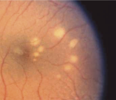
Figure 1.
Color photograph of the ocular fundus showing lesions (white spots) induced by blue-light exposures. There were two series of increasing energy with five exposures in each series. One series formed an inner arc of spots in the fovea at the edge of the macular pigment peak, whereas the outer arc of spots were in the parafovea in an area where macular pigment was optically undetectable. Lesions in the inner arc were smaller and there was no lesion at the location where the lowest energy was delivered. This image is from a control monkey with typical macular pigment.

