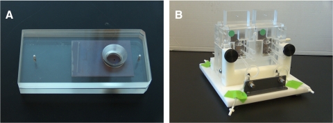
Figure 1.
Experimental setup. (A) Isolated BM/Ch was placed between two pieces of x-ray film such that it filled a 1.5-mm-diameter hole. The film sandwich was placed between two unbreakable polycarbonate plates (Lexan; Ledmark Industries, Lakewood, NJ) with a larger 120-mm hole. (B) Once the sandwich was assembled it was placed in a modified Ussing chamber. A mixture of proteins (ferritin, albumin, RNase A, and cytosine) dissolved in 4.5 mL of PBS-CM was added to the reservoir on one side of the tissue, with the opposite side containing 4.5 mL of PBS-CM only. Samples were collected from each reservoir after 4, 8, 16, 24, and 36 hours.

