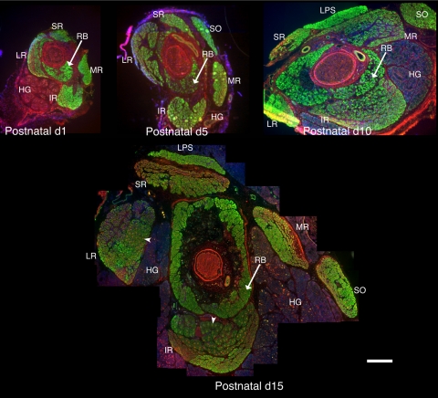
Figure 1.
Organization of the EOMs in postnatal developing orbits. Cryostat sections from the midbelly of developing orbits were stained with antibodies to sarcomeric α-actinin (green) and nmMyH IIB (red), and DAPI was used to stain the nuclei (blue). The major changes that occurred from P1 to P10 were an increase in the cross-sectional area of the Harderian gland and an increase in fiber size throughout the EOMs. Between P10 and P15, the distinction between the global and the orbital layers became pronounced, with obvious differences in the fiber size between the two layers and a distinct separation between the layers. Arrowheads in the P15 orbit indicate the fibers exhibiting the sarcomeric distribution of nmMyH IIB. RB, retractor bulbi; MR, medial rectus; LR, lateral rectus; SR, superior rectus, IR, inferior rectus; LPS, levator palpebrae superior; HG, Harderian gland. Scale bar, 500 μm.

