Figure 2.
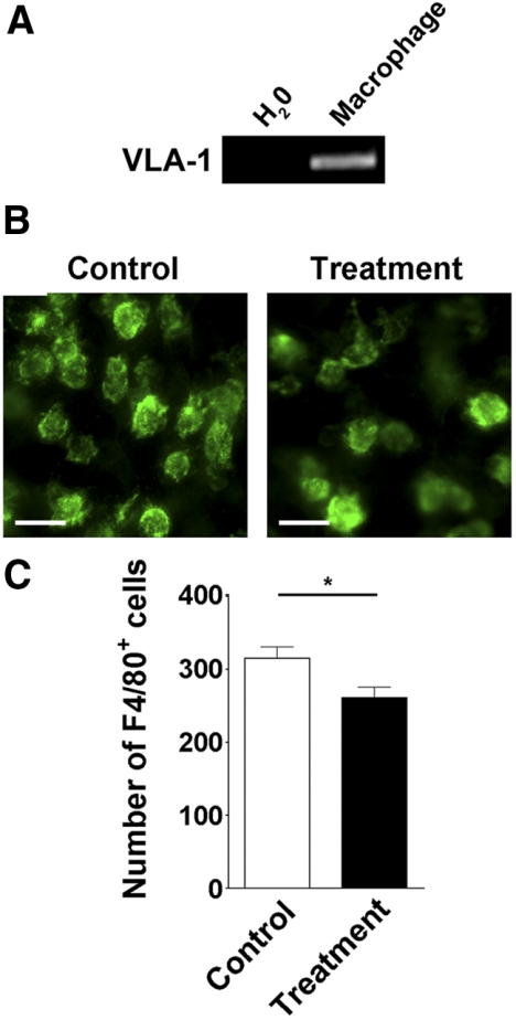
Corneal macrophage infiltration is inhibited by VLA-1 blockade. (A) RT-PCR analysis showing VLA-1 expression in corneal macrophages. (B) Representative images of immunofluorescence microscopic assays showing the number of F4/80-positive macrophages was significantly reduced in the cornea after VLA-1 antibody treatment. Green: F4/80. Scale bars, 25 μm. (C). Summarized data showing the difference of macrophage numbers between the VLA-1 antibody and isotype control treatment groups. *P < 0.05.

