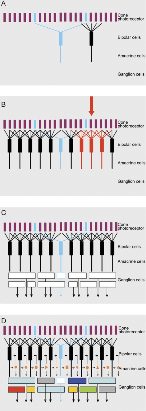
Figure 7A.
The four panels of this figure show exploded views of the retina's circuitry. Here, the mosaic of cone photoreceptors is shown at the top. (The late-evolving rod system is not shown, as it is a minor part of the retina's circuitry, and is used only in deep darkness.) The mosaic of cone photoreceptors is sampled by the “diffuse” bipolar cells (black), which receive input from all the cones within their reach, and by a small group of “blue bipolars,” which selectively contact the infrequent blue cones. This selectivity preserves the chromatic purity of the responses of the blue bipolar cells. Later retinal circuitry (not shown) compares the output of the blue (short wavelength) cones to that of the long wavelength cones. This comparison is the fundamental basis of mammalian color vision.
Figure 7B. The diffuse bipolar cells are far more numerous than the blue bipolars and come in 11 functional types, each transmitting a different component of the cone's output. Stimulation of an individual cone (red arrow) transmits information to all the bipolar cells that contact it (red bipolar cells in the illustration). In this case the red bipolar cells would all be of one functional type; for example, all of them would be ON-transient bipolars. But that same (arrow) cone would also be contacted by bipolar cells of the other 10 types, carrying different components of the cone's output. The whole array cannot be shown in a planar diagram like this one—there is not enough space for all the bipolar cells in a single plane, and 12 different colors of bipolar cell would be required.
Figure 7C. The different types of bipolar cells synapse on different types of ganglion cells. Since each type of bipolar cell conveys a different type of information to the inner retina, each conveys its own particular version of the visual input to the ganglion cell(s) on which it synapses. This is the initial step in the creation of ganglion cell diversity. In the example, the blue bipolar cell synapses on a specific type of ganglion cell, which then becomes a blue-sensitive ganglion cell. The same is true for each of the other bipolar cell types. If this were the whole story, the organization of the inner retina would be relatively simple. There might still be 12 (or more) functional types of ganglion cells, but the bipolar cells would provide the only drive to the ganglion cells, so that the ganglion cell response would be determined purely by the response tuning of the bipolar cell, the physiology of the bipolar cell synapse and the ganglion cell's ion channels.
Figure 7D. The final responses of the ganglion cells are highly selective: Some respond only to a particular direction of stimulus movement, some specifically to low or to high contrast, some specifically to rapid movement, and so on. This final diversity is created by the action of amacrine cells. Amacrine cells receive a direct excitatory input from bipolar cells, and they make inhibitory synaptic inputs back onto the axon terminals of the bipolar cells. This is a classic feedback system, creating a control point at the bottleneck where information enters the inner retina from the outer retina. However, amacrine cells also synapse on each other; and they feed forward on retinal ganglion cells. These feed-forward synapses are responsible for some of the more interesting properties of the ganglion cell response. A famous case is the starburst amacrine cell, which creates direction selectivity in one type of retinal ganglion cell.46,47 Not shown are the wide-field amacrine cells, which can span the whole width of the retinal surface.

