Figure 4.
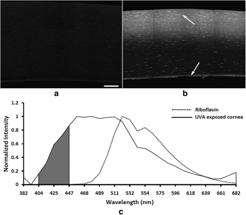
(a) Collagen autofluorescence image (400–450 nm) of control corneas treated with riboflavin. Corneas were fixed and washed to remove excess riboflavin. (b) Collagen autofluorescence image (400–450 nm) of a riboflavin-soaked and 30-minute UVA-exposed cornea showing increased fluorescence in the anterior corneal stroma after removal of the riboflavin. Note the presence of cellular autofluorescence in the corneal stroma and corneal endothelium (arrows). (c) Emission spectra of nonlinear optical signals generated by 760-nm femtosecond laser light from riboflavin and corneas after UVA-induced CXL. Riboflavin showed peak fluorescence at 521 nm, whereas CXL corneas showed peak fluorescence at 425 nm. Bar, 100 μm.

