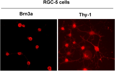
Figure 1.
RGC-5 cells expressed RGC markers. RGC-5 cells were plated in eight-well chamber slides, treated overnight with 2.0 μM SS, and immunostained with antibodies against Brn3a and Thy-1. The results showed that the RGC-5 cells differentiated after SS treatment and expressed Brn3a and Thy-1, markers characteristic of RGCs. Magnification, ×40.

