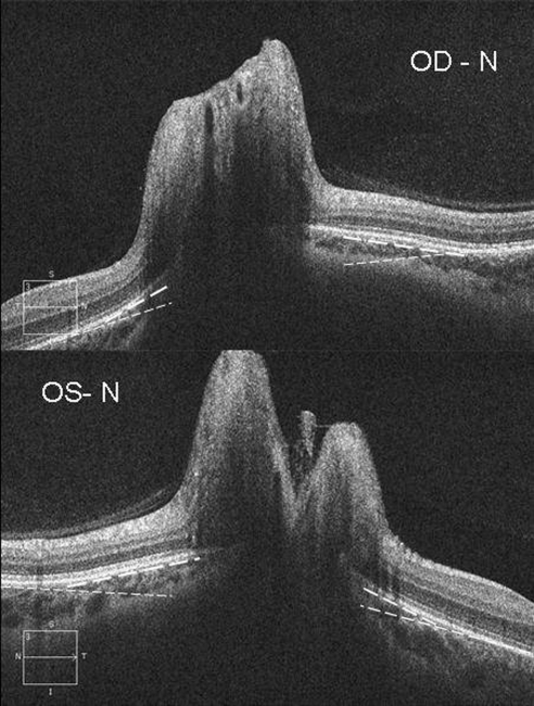
Figure 1.
The right (OD, top) and left (OS, bottom) eye from one patient with papilledema showing inward positive angulation (toward the vitreous) of the peripapillary RPE/BM layer relative to the more peripheral peripapillary regions of the retina (broken lines). Note the angle on the nasal side (N) of the neural canal is greater than on the temporal angle for each eye.

