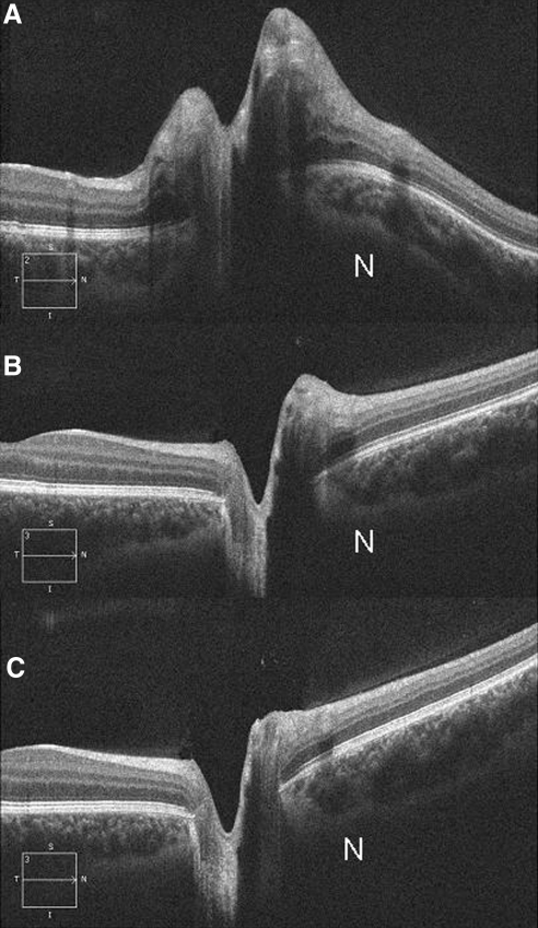
Figure 2.
Sequential HD-OCT of the right eye from a patient with papilledema followed for a period of 12 weeks. (A) Photograph obtained at baseline showing marked elevation of the optic disc, thickening of the RNFL, and inward angulation of the temporal RPE/BM at the margin and severe inward deformation and displacement of the nasal RPE/BM. Note that the sclera/choroid are also deflected inward (worse at nasal border [N]). (B) Photograph obtained 8 weeks after a 15-lb weight loss revealing a significant decrease in disc elevation, RNFL thickening, and straightening of the RPE/BM layer. (C) Photograph obtained at 12 weeks after a 24-lb weight loss reveals resolution of papilledema with a negative RPE/BM. At each time point, the left eye (not shown) had similar findings.

