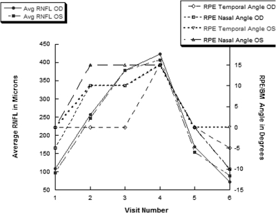
Figure 4.
For our prototypical case, the average RNFL and RPE/BM angles at neural canal borders changed in parallel over six visits. The RPE/BM angles became positive as the RNFL swelling increased. Time point five is 1 week after a ventricular shunt was placed to normalize the intracranial hypertension and reveals normalization of the RPE/BM angles in both eyes.

