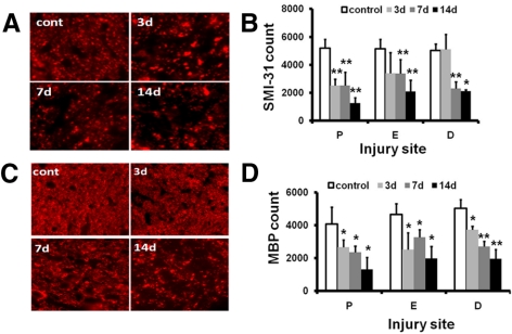
Figure 4.
Immunohistochemistry of SMI-31 and MBP of the control and the crush-injured optic nerves. (A) SMI-31 of proximal optic nerve cross-sections from control (cont) and ONC injury mice at 3, 7, and 14 days. (B) Quantitative SMI-31–positive axon counts of the optic nerve at proximal (P), crushed epicenter (E), and distal (D) sites. (C) MBP of the proximal optic nerve cross-sections from the control (cont) and ONC-injured mice at 3, 7, and 14 days after injury. (D) Quantitative MBP-positive axon counts at the three selected sites. The timing of myelin injury reflected as loss of MBP-positive axon counts is similar to that of SMI-31. *P < 0.05, **P < 0.01. Magnification, 60×.

