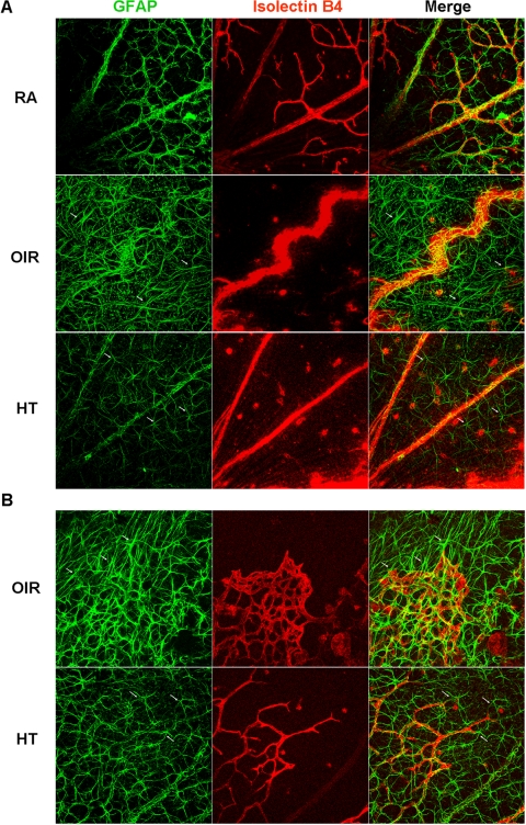
Figure 4.
HT improves the status of astrocyte network and morphology. OIR mice were treated with hyperoxia (75% oxygen; HT) or were maintained in room air (OIR) from P14 to P17. Mice maintained in room air (RA) from P1 to P17 are control. Retinal flatmounts were stained with isolectin B4 (red) and anti-GFAP (green). Representative confocal images are shown (n = 9 retinas from 9 mice). (arrows) Examples of astrocytes with different morphology in OIR (spindle-shaped) and HT (stellate). The punctate staining for GFAP in the flatmounts represents cross-sections of Müller cells expressing GFAP. Original magnification, ×20. (A) Central retina. (B) Revascularization area.

