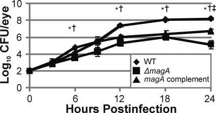
Figure 2.
Intraocular growth of K. pneumoniae during experimental endophthalmitis. Eyes were injected with 100 CFU of wild-type, ΔmagA, or magA complement K. pneumoniae and were enucleated at the indicated time points, homogenized, and plated on BHI agar for bacterial quantification. *P ≤ 0.05 for wild-type versus ΔmagA. †P ≤ 0.05 for wild-type versus magA complement. ‡P ≤ 0.05 for ΔmagA versus magA complement. Two-tailed, two-sample t-tests assuming equal variance were used to statistically compare groups.

