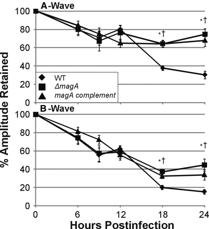
Figure 3.
Retinal function during experimental K. pneumoniae endophthalmitis. Eyes were injected with 100 CFU of wild-type, ΔmagA, or magA complement K. pneumoniae and were dark adapted at least 6 hours before electroretinography. Infected eyes were compared with the contralateral mock-injected or absolute control eye and were reported at percentage of amplitude of A-wave or B-wave retained. *P ≤ 0.05 for wild-type versus ΔmagA. †P ≤ 0.05 for wild-type versus magA complement. Two-tailed, two-sample t-tests assuming equal variance were used to statistically compare groups.

