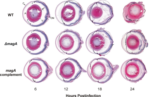
Figure 5.
Histology during experimental K. pneumoniae endophthalmitis. Eyes were injected with 100 CFU of wild-type, ΔmagA, or magA complement K. pneumoniae and were enucleated at the indicated time points postinfection, processed for histology, and stained with hematoxylin and eosin. Eyes at 6 hours for all strains were indistinguishable from mock-injected or absolute control eyes at any time point. Images are magnified approximately 10-fold. A, aqueous humor; C, cornea; L, lens; ON, optic nerve; R, retina; V, vitreous.

