Figure 2.
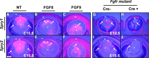
Spry1 and -2 expression in FGF transgenic and FGFR mutant mice. In situ hybridization with 35S-labeled Spry1 and -2 riboprobes was performed on sections of nontransgenic (NT) (A, D), FGF8 (B, E), and FGF9 (C, F) transgenic and FGFR mutant (G–J) embryos. Spry1 and -2 were upregulated at the transition zone in the nontransgenic lenses (A, D, arrows). Lens fiber–specific expression of FGF8 (B, E) or FGF9 (C, F), weakly induced Spry1 (B, C, arrows), and strongly induced Spry2 (E, F, arrows) in the lens epithelial cells. (B–F, dashed lines) The lens. Spry1 (H, arrow) and Spry2 (J, arrow) expression was reduced in FGFR mutant lenses in contrast to Cre− controls (G, I). Staining within the lens core (*) in (A–F) and in the RPE (G) are artifacts of dark-field illumination. True hybridization signals appeared as dots (arrows), and diffraction artifacts had a hazy, less-defined appearance that was visible even in the complete absence of a signal. cs, corneal stroma; le, lens epithelium; lf, lens fibers; r, retina. Scale bar, 40 μm.

