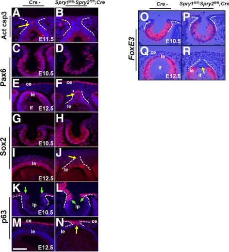
Figure 7.
Early lens differentiation in Spry mutant embryos. Immunohistochemistry (A–N) and in situ hybridizations (O–R) were performed on control (Cre−) and Spry mutant embryos, to detect expression of activated caspase 3 (A, B), Pax6 (C–F), Sox2 (G–J), p63 (K–N), and FoxE3 (O–R). In situ hybridizations were performed using 35S-labeled riboprobes (O–R). Activated caspase 3 was seen in the stalks of Cre− controls (A, arrow) but not in Spry mutants. Pax6 (D, F) and Sox2 (H, J) were expressed in their normal spatial pattern in Spry mutants. Spry mutant lens stalks, however, showed reduced Sox2 expression compared to lens epithelial cells (J, arrow). p63, normally excluded from the lens placodal cells (K, green arrows), was expressed at the anterior margins of the lens pit (L, arrows) and in the stalks (N, arrow) of Spry mutants. FoxE3 expression in the invaginating lens placodal cells at E10.5 (O) and epithelial cells (Q) was similar to controls (O). Spry mutant stalk cells did not express FoxE3 (R, arrow). ce, corneal epithelium; le, lens epithelium; lf, lens fibers; lp, lens pit. Scale bar in (M): (A–D, M, N) 10 μm; (E, F, I–L, O–R) 15 μm.

