Figure 6.
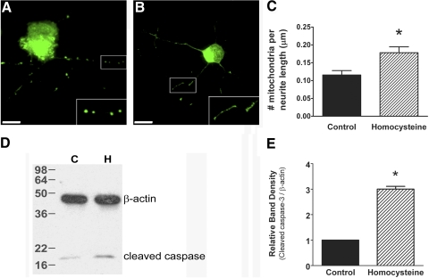
Exposure of primary ganglion cells to 50 μM homocysteine induces mitochondria that are smaller and more numerous and increases levels of cleaved caspase-3. Representative images of primary ganglion cells loaded with dye; no treatment (A) versus 50 μM homocysteine (B). Scale bar, 10 μM. (inset) Higher magnification of boxed area. Quantification of the number of mitochondria per length of neurite (C) shows that mitochondrial number is higher in primary ganglion cell neurites after treatment with 50 μM homocysteine (no treatment, 0.1156 ± 0.012, 50 μM; homocysteine, 0.1781 ± 0.017; P < 0.016). Immunoblot analysis performed on protein isolated from primary ganglion cells after exposure to 50 μM homocysteine revealed higher levels of cleaved caspase-3 than in control primary ganglion cells (C, control; H, homocysteine-treated) (D). Densitometric analysis of cleaved caspase-3. (E) Control, 1.00 ± 0.00; homocysteine-treated, 3.00 ± 0.11; P < 0.003.

