Figure 5.
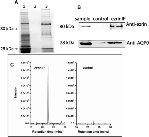
Coimmunoprecipitation of AQP0 with ezrin antibody. Lens fiber cell WIF was extracted with 25 mM tris, 150 mM NaCl, 5 mM EDTA, 1% Triton X-100, 1 mM PMSF, 0.1% SDS, pH 7.4 (lane 1) and immunoprecipitated with anti-ezrin antibody (lane 3). The control sample was treated in the same way except without adding anti-ezrin antibody (lane 2). The samples were separated on SDS-PAGE and followed by Coomassie blue stain (A) and Western blotting (B). Bands at 28 and 80 kDa were detected by Coomassie blue stain in the immunoprecipitate using the ezrin antibody. The presence of ezrin and AQP0 were confirmed by Western blotting with anti-ezrin and anti-AQP0 antibodies and LC-MS/MS. Weak ezrin and AQP0 signals were also detected in the control sample, but the signals of these proteins in immunoprecipitated sample were significantly higher than in the control sample. The selected ion chromatograms of AQP0 peptide 239 to 259 (m/z 733.5) in the control sample and immunoprecipitated sample are plotted (C).

