Figure 1.
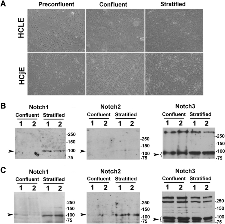
Notch receptors in human corneal and conjunctival epithelial cell cultures. (A) Representative phase contrast micrographs showing HCLE and HCjE cells at three stages of culture: preconfluent, confluent in the absence of serum, and stratified after 7 days of serum supplementation. (B) By Western blot analyses, the intracellular domain of Notch3, but not Notch1 or Notch2, was present in undifferentiated confluent HCLE cells. The Notch3 antibody bound to a major band in the 80 to 90 kDa region corresponding to the intracellular domain, as previously described.14 Induction of cell differentiation and stratification resulted in binding of the Notch1 antibody to the 100 kDa intracellular domain. Weak binding of the Notch2 antibody was observed after induction of cell differentiation. (C) The intracellular domains of Notch1 to Notch3 were detected in HCjE cells, primarily after induction of cell differentiation and stratification. Experiments were performed in duplicate. Sample lanes were loaded with 50 μg (Notch1), 100 μg (Notch2), and 20 μg (Notch3) total protein. Arrowheads indicate the position of the Notch transmembrane intracellular domain.

