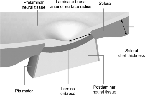
Figure 2.
Model geometry. Five tissue regions were modeled: corneoscleral shell, lamina cribrosa (LC), prelaminar neural tissue (PLNT, including the retina and choroid), postlaminar neural tissue (ON, including the optic nerve), and pia mater. IOP was represented as a homogeneous force on the interior surfaces. The apex of the region representing the cornea was constrained in all directions to prevent displacement or rotation. See Table 1 for the factor ranges. Scleral thickness was parameterized over the shell, such that the scleral thickness at the canal wall remained unchanged.

