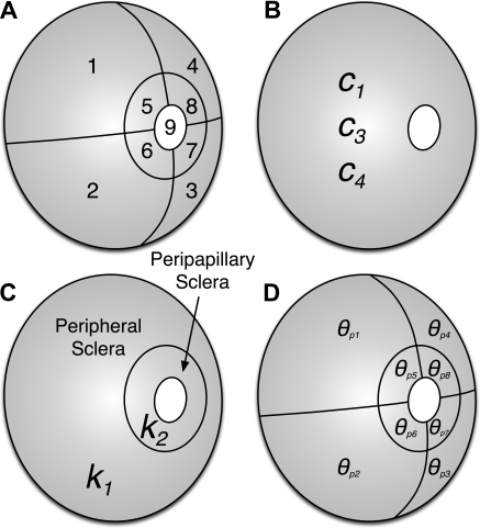
Figure 3.
(A) Thirteen unique model parameters are estimated for each eye. Each reconstructed posterior scleral geometry was subdivided into nine regions: regions 1 to 4 represent the peripheral sclera, regions 5 to 8 the peripapillary sclera, and region 9 the ONH. (B) Three stiffness parameters were uniformly attributed to the entire scleral shell (c1, first Mooney-Rivlin coefficient; c3, exponential fiber stress coefficient; c4, uncrimping rate of the collagen fibers). (C) Two structural parameters—fiber concentration factors k1 and k2—were attributed to the peripapillary and peripheral sclera respectively. (D) Eight other structural parameters—preferred fiber orientations θp1 to θp8—were attributed to each of the eight scleral regions respectively. Note that the ONH was assumed linear isotropic with an elastic modulus fixed to 1 MPa for all normal and glaucomatous monkey eyes. This assumption is reasonable, as the ONH has been shown to have little impact on IOP-induced scleral deformations.9,46

